


Update on Retinal Imaging: Advances Transforming Clinical Practice
Overview
Recent advancements in retinal imaging technologies, including home OCT devices, swept-source OCT, OCT-angiography, and AI integration, are revolutionizing early detection and management of retinal diseases. These innovations enhance imaging speed, resolution, and accessibility, enabling personalized patient care and remote monitoring.
Background
Retinal imaging is central to ophthalmology, providing critical insights into retinal health and disease. Traditional modalities like fluorescein angiography remain important, but newer technologies such as OCT and OCT-angiography offer non-invasive, high-resolution visualization of retinal structures and vasculature. The integration of artificial intelligence and home-based imaging systems is expanding the scope of disease monitoring beyond clinical settings, improving early detection and treatment outcomes.
Data Highlights
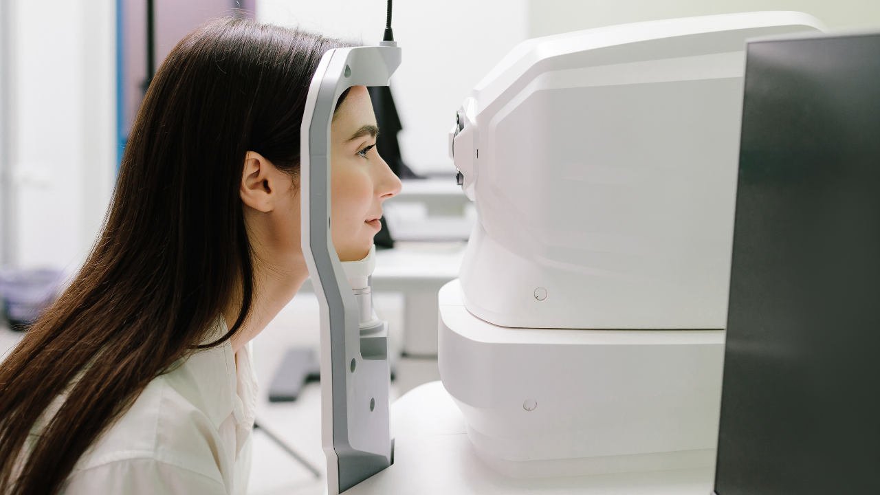
The FDA recently authorized the Notal Vision Home OCT system, the first home-use OCT device cleared for monitoring chronic retinal conditions like neovascular age-related macular degeneration (nAMD). Scan speeds in advanced OCT systems now exceed 150 kHz, significantly improving workflow efficiency and image quality. Adaptive optics platforms such as Imagine Eyes’ RTX1 are approved in select countries, offering cellular-level imaging capabilities. AI-powered software automates segmentation and quantification of retinal features, enhancing diagnostic accuracy and reducing clinician workload.
Key Findings
- The FDA’s De Novo clearance of the Notal Vision Home OCT marks a regulatory milestone, enabling remote monitoring of chronic retinal diseases and reducing in-office visit frequency.
- Swept-Source OCT provides deeper, clearer imaging of the choroid and vitreoretinal interface, aiding diagnosis of complex conditions like pachychoroid disorders and advanced glaucoma.
- OCT-Angiography offers a non-invasive alternative to dye-based angiography, with improved motion correction and segmentation enhancing vascular imaging reliability.
- Fundus Autofluorescence imaging, especially near-infrared FAF, complements other modalities by assessing retinal pigment epithelium health and deeper retinal structures.
- Wide-field imaging systems now integrate with OCT and angiography, capturing high-resolution peripheral retinal images critical for managing diabetic retinopathy and retinal detachments.
- Artificial intelligence tools assist in early disease detection and monitoring by automating image analysis, though they require diverse training datasets for optimal performance.
Clinical Implications
Clinicians can leverage home OCT devices to monitor disease progression remotely, improving patient convenience and enabling earlier intervention. The adoption of swept-source OCT and OCT-A enhances diagnostic capabilities for complex retinal diseases, while AI integration streamlines image analysis and supports clinical decision-making. Incorporating wide-field and autofluorescence imaging provides a comprehensive assessment of retinal health, facilitating tailored treatment strategies.
Conclusion
The evolving landscape of retinal imaging, characterized by technological innovation and AI integration, is transforming patient care by enabling earlier detection, precise monitoring, and personalized management of retinal diseases. These advancements promise to improve clinical outcomes and expand access to high-quality retinal diagnostics.
Related Resources & Content
- Update on Retinal Imaging -- Article Source 2024
This content is an AI-generated, fully rewritten summary based on a published scholarly article. It does not reproduce the original text and is not a substitute for the original publication. Readers are encouraged to consult the source for full context, data, and methodology.